
Abnormal RNS of the trapezius is 100% specific for ALS as opposed to CSA, whereas absence of decrement in the deltoid, in patients with upper limb onset, may exclude ALS. Among 53 ALS and 37 CSA patients, RNS of the abductor pollicis brevis, upper trapezius, and deltoid muscles revealed an abnormal decremental response in 31%, 51%, and 75% of the ALS patients, but only in 3%, 0%, and 20% of the CSA patients, respectively. Repetitive nerve stimulation (RNS) studies may help differentiate between them. COMMENTARYĬervical spondylotic amyotrophy (CSA), presenting with regional muscle atrophy, muscle weakness, and minimal long tract signs, often requires clinical differentiation from ALS. Studying six upper and five lower extremity muscles offered a > 98% positive yield, and beginning with a proximal muscle in the arm and leg, such as the deltoid and the quadriceps, respectively, and then examining a distal muscle, is recommended. Abnormalities also were more likely to be found in muscles of the initially affected limb compared to limbs subsequently affected. In the arm, these muscles included the first dorsal interosseous, the abductor pollicis brevis, and the abductor digiti minimi, and, in the leg, the tibialis anterior, the medial gastrocnemius, and the tibialis posterior/flexor digitorum longus. EMG findings from 5,315 muscles examined in these patients revealed that regardless of where the first signs of disease occurred, distal limb muscles were the most sensitive in providing evidence of lower motor neuron dysfunction, in the form of active and chronic denervation.

1 Mean age was 60.5 years, 89% were Caucasian, 5% were African American, and the male to female ratio was 1:2. Statistical analysis encompassed the Fisher exact test and pair-wise comparison of proportions.Īmong 617 screened patients, 354 met eligibility criteria, including 64 with definite ALS, 204 with probable ALS, and 86 with clinically probable, laboratory-supported ALS, as defined by consensus guidelines. Reanalysis of EMG findings using Awaji criteria, allowing fasciculation potentials to serve as evidence of active denervation when chronic denervation is concomitantly present, also was undertaken. Patients with mononeuropathies were included, but muscles innervated by the affected nerves were excluded from analysis. To avoid possible neuropathic confounding influences, patients aged 60-75 years with absent sural sensory responses on nerve conduction studies also were excluded, even in the absence of clinical polyneuropathy. Patients with underlying sensorimotor polyneuropathy, or cervical or lumbosacral spondylomyelopathy were excluded. 31, 2012, with a clinical diagnosis of ALS and who underwent EMG testing. To answer the above question, Babu et al performed a retrospective chart review of 617 consecutive patients, seen at the Cleveland Clinic between Jan. Given the innate discomfort of EMG, which muscles and combination of muscles might offer the highest yield, allowing testing to be minimized as much as possible? In each of the cervical and lumbosacral regions, at least two muscles innervated by different roots and different peripheral nerves must have positive sharp waves or fibrillation potentials.
Als emg findings plus#
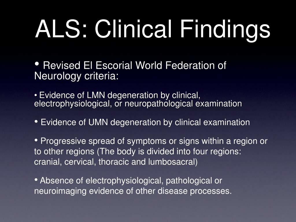
Revised El Escorial ALS diagnostic criteria require evidence of active and chronic denervation, the latter defined as large, unstable, polyphasic motor unit potentials of increased duration with a reduced interference pattern in cervical, thoracic, and lumbar regions or in bulbar muscles plus two spinal regions. Muscle Nerve 2017:56:36-44.Īlthough electrodiagnostic findings of active denervation on needle electromyography (EMG), comprising positive sharp waves and fibrillation potentials, are not pathognomonic for amyotrophic lateral sclerosis (ALS) and may be seen in any denervating process, these abnormalities are the sine qua non for making a diagnosis of ALS in the appropriate clinical setting. Optimizing muscle selection for electromyography in amyotrophic lateral sclerosis. SYNOPSIS: In the appropriate clinical setting, electromyography studies of multiple, distal muscle groups in three separate spinal regions showing acute and chronic denervation, are pathognomonic for the diagnosis of amyotrophic lateral sclerosis. Rubin reports no financial relationships relevant to this field of study.

Professor of Clinical Neurology, Weill Cornell Medical Collegeĭr.


 0 kommentar(er)
